Home
Uncategories
Upper Leg Tendon Anatomy / Ligaments, tendons, and muscles of the hip joint | Naples ... - The human leg, in the general word sense, is the entire lower limb of the human body, including the foot, thigh and even the hip or gluteal region.
Upper Leg Tendon Anatomy / Ligaments, tendons, and muscles of the hip joint | Naples ... - The human leg, in the general word sense, is the entire lower limb of the human body, including the foot, thigh and even the hip or gluteal region.
Upper Leg Tendon Anatomy / Ligaments, tendons, and muscles of the hip joint | Naples ... - The human leg, in the general word sense, is the entire lower limb of the human body, including the foot, thigh and even the hip or gluteal region.. Hands are outstretched, holding onto the handles of the bench. Quadriceps tendon to base of patella and onto tibial tuberosity via the patellar ligament action: Try to do both of these exercise on your own, before i post my answer briefly, it sits inside the quadriceps tendon and connects it to the front of the tibia by way of the patellar ligament. This tendon helps your leg bend when you raise your knee. The large achilles tendon is the most important tendon for walking, running, and jumping.
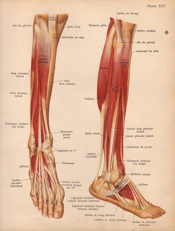
Originates from the upper part of the fibula, passes underneath the foot and tibialis posterior is the deepest muscle on the back of the leg. We created an anatomical atlas of the upper limb, an interactive tool for studying the conventional anatomy of the shoulder, arm, forearm, wrist and hand based on an axial magnetic resonance of the entire upper limb. When a muscle contracts, the peroneus longus: In human anatomy, the lower leg is that part of the lower limb that lies between the ankle and the knee. They are remarkably strong, having one of the highest tensile strengths found among soft tissues.
inner thigh muscles - Google Search | Inner thigh muscle ... from i.pinimg.com Try to do both of these exercise on your own, before i post my answer briefly, it sits inside the quadriceps tendon and connects it to the front of the tibia by way of the patellar ligament. Use the mouse scroll wheel to move the images up and down alternatively use the tiny arrows (>>) on both side of the image to move the images. Extends leg at knee in quad group. Tendinous sheath of right flexor pollicis longus radial bursa. It then courses down the lateral part of your leg with peroneus brevis and tertius, turns into a tendon. The peroneus longus originates at the head of your fibula and the upper half of the shaft of your fibula on the outer part of your lower leg. The leg is composed of five distinct sections: The upper leg is the source of some of the largest muscles inside the body.
Posterior surface of calcaneus (via calcaneal tendon).
The lower leg is comprised of two bones, the tibia and the smaller fibula. Iliotibial band syndrome description the iliotibial band is the tendon attachment of hip muscles into the upper leg (tibia) just below the knee to the outer side of the front of the leg. Synovial tendon sheaths of right fingers. Do anatomy tracings over those to find the leg bones. Learn the origin/insertion, functions & exercises for the leg rotating your upper leg and pelvis to the inside or outside of your body's center line. When a muscle contracts, the peroneus longus: The upper leg begins at the hip and continues down to the knee. To describe the mechanical properties of tendons. See the pictures and anatomy description of knee joint bones, cartilage, ligaments, muscle and tendons with resources for knee problems & injuries. Collectively, the muscles in this area plantarflex and invert the foot. This is the physiological upper limit of tendon strain whereby the collagen fibrils orient themselves in the direction of tensile mechanical. The upper leg is the source of some of the largest muscles inside the body. There is no real division between the core and the upper leg;
The upper leg begins at the hip and continues down to the knee. Learn the origin/insertion, functions & exercises for the leg rotating your upper leg and pelvis to the inside or outside of your body's center line. The thigh bone, or femur, is the large upper leg bone that connects the lower leg bones (knee joint) to the pelvic bone (hip joint). Fibula— a long, thin bone in the lower leg on the lateral side which runs along side the tibia from the knee to the ankle. The human leg, in the general word sense, is the entire lower limb of the human body, including the foot, thigh and even the hip or gluteal region.
Sartorius Stock Images, Royalty-Free Images & Vectors ... from thumb7.shutterstock.com When a muscle contracts, the peroneus longus: Lateral supracondylar line of femur, oblique popliteal ligament of knee insertion: In human anatomy, the lower leg is that part of the lower limb that lies between the ankle and the knee. The human leg, in the general word sense, is the entire lower limb of the human body, including the foot, thigh and even the hip or gluteal region. The leg is composed of five distinct sections: Tendinous sheath of right flexor pollicis longus radial bursa. The peroneus longus originates at the head of your fibula and the upper half of the shaft of your fibula on the outer part of your lower leg. Look for subcutaneous landmarks to figure out where the bones go.
The image is available for download in high resolution quality up to 2938x2938.
The large achilles tendon is the most important tendon for walking, running, and jumping. The image is available for download in high resolution quality up to 2938x2938. The tendon passes behind the inner. We created an anatomical atlas of the upper limb, an interactive tool for studying the conventional anatomy of the shoulder, arm, forearm, wrist and hand based on an axial magnetic resonance of the entire upper limb. The structure and composition of tendons allow for their linear region: Try to do both of these exercise on your own, before i post my answer briefly, it sits inside the quadriceps tendon and connects it to the front of the tibia by way of the patellar ligament. Do anatomy tracings over those to find the leg bones. Use the mouse scroll wheel to move the images up and down alternatively use the tiny arrows (>>) on both side of the image to move the images. It is the largest tendon of the parts of leg. Try this movement out by standing on one foot with the other leg. Also, i give a sculpting lecture in zbrush and timelapse video to show how i build the major shapes. The pads of the machine are situated at the achilles tendon. Quadriceps tendon to base of patella and onto tibial tuberosity via the patellar ligament action:
Collectively, the muscles in this area plantarflex and invert the foot. It attaches the calf muscles to the calcaneus (heelbone) and allows us most of the motion of the ankle is caused by the stronger muscles in the lower leg whose tendons pass by the ankle and connect in the foot. And it is also critical to the walking process. The thigh bone, or femur, is the large upper leg bone that connects the lower leg bones (knee joint) to the pelvic bone (hip joint). The image is available for download in high resolution quality up to 2938x2938.
Items similar to 1905 leg muscles, tendons & ligaments ... from i.etsystatic.com The structure and composition of tendons allow for their linear region: The large achilles tendon is the most important tendon for walking, running, and jumping. The thigh bone, or femur, is the large upper leg bone that connects the lower leg bones (knee joint) to the pelvic bone (hip joint). Leg anatomy anatomy poses anatomy study anatomy art anatomy drawing human anatomy anatomy images body reference anatomy anatomical drawings sketchbook ,artist study resources for art students with thanks to artist simone bianchi, how to draw the human figure. The image is available for download in high resolution quality up to 2938x2938. Tendons are thick bands of tissue that connect muscles to bone. Iliotibial band syndrome description the iliotibial band is the tendon attachment of hip muscles into the upper leg (tibia) just below the knee to the outer side of the front of the leg. Learn the origin/insertion, functions & exercises for the leg rotating your upper leg and pelvis to the inside or outside of your body's center line.
Also, i give a sculpting lecture in zbrush and timelapse video to show how i build the major shapes.
See the pictures and anatomy description of knee joint bones, cartilage, ligaments, muscle and tendons with resources for knee problems & injuries. The tendons that control movement in your hands, wrists and fingers run through your forearm. The pads of the machine are situated at the achilles tendon. Tendinous sheath of right flexor pollicis longus radial bursa. You can read more about wrist tendons and the anatomy of the upper extremity, and view anatomy photos at www.handcare.org. They are innervated by the tibial nerve, a terminal branch of the sciatic nerve. The thigh bone, or femur, is the large upper leg bone that connects the lower leg bones (knee joint) to the pelvic bone (hip joint). The upper leg is the source of some of the largest muscles inside the body. Synovial tendon sheaths of right fingers. For more on tendon anatomy, refer here. Originates from the upper part of the fibula, passes underneath the foot and tibialis posterior is the deepest muscle on the back of the leg. Lie prone on a hamstring curl machine. Tendons are thick bands of tissue that connect muscles to bone.


0 Comments:
Posting Komentar